
Highlights
- Established mouse myocardial infarction model followed longitudinally with ¹⁸F-FDG PET/CT on BIOEMTECH β-eye™
- Three time points post-surgery (Days 7, 14, 28) with consistent acquisition and post-processing conditions
- PET findings supported by ECG at Day 28 (ST-segment depression and ectopic heartbeats in ischaemic mice)
Collaboration: Study was conducted in collaboration with Iordanis Mourouzis and Konstantinos Patakioutis from the Department of Pharmacology, Medical School, National and Kapodistrian University of Athens
Introduction
Myocardial infarction has been extensively investigated in preclinical cardiovascular research using a range of experimental models. However, improved and more accurate therapies remain an unmet need and require further evaluation.
Here, we assessed an established experimental model using non-invasive PET/CT imaging with a twofold objective:
- to further investigate and evaluate myocardial metabolic activity in both healthy and ischaemic myocardium following surgical induction, and
- to develop a robust preclinical imaging platform capable of objectively assessing the efficacy of emerging therapeutic strategies.
Study design
The study included eight C57BL/6 mice, with ischaemic (myocardial infarction) (n=4) and control (healthy) (n=4) age-matched groups, imaged on the BIOEMTECH β-eye™ system.
Imaging protocol
Each imaging session followed an intravenous injection of 6 MBq ¹⁸F-FDG. PET acquisition was initiated 80 minutes post-injection and was followed by a CT scan for anatomical localization.
Methods
A surgical protocol for induction of myocardial ischaemia was performed in male C57BL/6 mice (10 weeks old). A skin and muscle incision was made at the fourth intercostal space. Following pericardiotomy, the left coronary artery was ligated using an 8-0 silk round-bodied suture.
On day 6 post-surgery, ultrasonographic evaluation revealed a significant reduction in myocardial functionality due to ischaemia. On days 7, 14, and 28 post-surgery, animals were anaesthetised and received an intravenous administration of 6 MBq ¹⁸F-FDG. Eighty minutes later, animals underwent high-resolution PET (BIOEMTECH β-eye™) followed by CT scanning. On day 28, an electrocardiographic evaluation of the myocardium’s conduction system was performed.
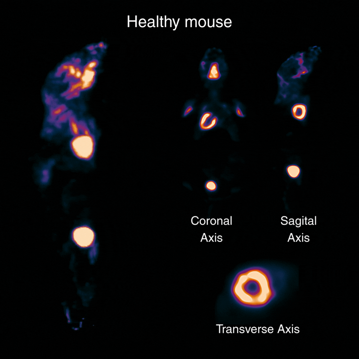
Figure 1: ¹⁸F-FDG PET imaging of a healthy C57BL/6 mouse, acquired 80 minutes post-injection with a 20-minute scan following administration of 6 MBq, shown in coronal, sagittal and transverse views.
Results
PET/CT signal acquisition from viable myocardium confirmed increased ¹⁸F-FDG uptake in inflamed viable myocardium, whereas the ischaemic region exhibited decreased uptake, as shown in sagittal, transverse and coronal views (Figure 2), particularly when compared to control animals (Figure 1). This reduction was predominantly evident in the apical region of the heart.
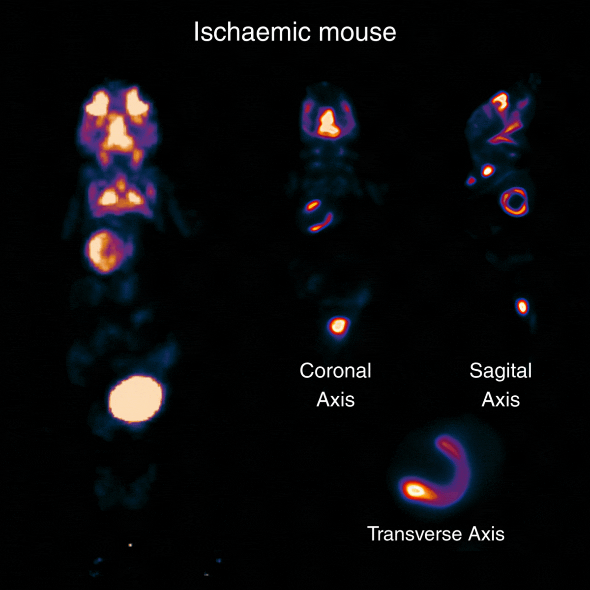
Figure 2: ¹⁸F-FDG PET imaging of an ischaemic C57BL/6 mouse at Day 30 post-surgery, acquired 80 minutes post-injection with a 20-minute scan following administration of 6 MBq, shown in coronal, sagittal and transverse views.
On days 14 and 28, imaging findings were comparable to those observed on day 7 post-surgery. In both groups, the same colour scale was applied during post-processing.
Electrocardiography showed normal recordings in control mice, whereas ischaemic mice exhibited ST-segment depression and ectopic heartbeats (Figure 3).
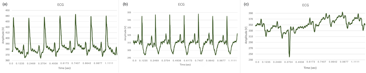
Figure 3: ECG recordings of C57BL/6 mice
panel a Non-ischaemic mice: sinus rhythm, panel b Ischaemic mice: ST-segment depression, panel c Ischaemic mice: arrhythmia, ectopic heartbeat.
Image processing and quantification
To convert imaging data into quantitative endpoints, analysis was performed using Imalytics, enabling:
- co-registration of PET and CT datasets (Figure 4),
- segmentation of myocardial regions of interest,
- quantification of ¹⁸F-FDG uptake, and
- comparison of metabolic activity between healthy and ischaemic myocardial regions across time points.
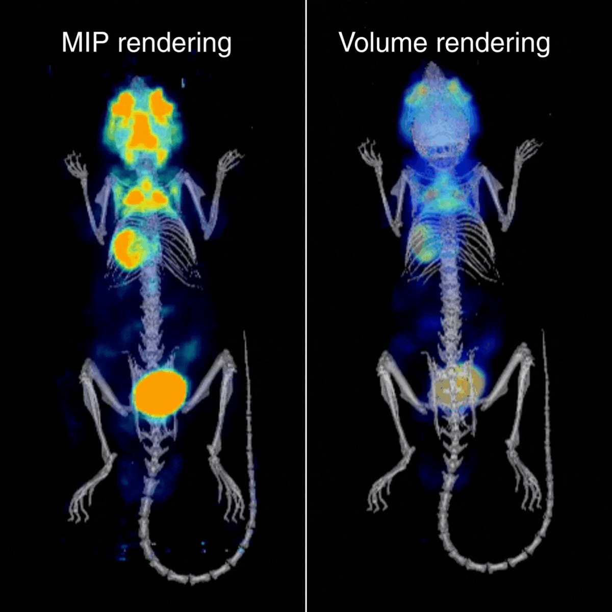
Figure 4: Maximum-intensity projection (MIP) and 3D volume renderings of the ¹⁸F-FDG PET/CT dataset, illustrating whole-body tracer distribution and supporting visual assessment alongside quantitative analysis.
This workflow supported an objective assessment of evolving metabolic differences, contributed to validation of the infarct model, and provides a foundation for non-invasive evaluation of novel cardioprotective drugs.
Conclusion
These results provide qualitative functional evidence of ischaemic lesions in this animal model. They could further be used to deliver quantitative data when combined with superimposed CT scans and appropriate placement of regions of interest on the myocardium (whole heart, apex, and base of the heart), offering a valuable, non-invasive, objective, and accurate approach in preclinical cardiovascular research.
In addition, electrocardiographic findings, specifically ST-segment depression and ectopic heartbeats in ischaemic subjects, were consistent with the results obtained from high-resolution PET imaging.