
Highlights
- Fast whole-body planar imaging for efficient preclinical in vivo screening
- Representative applications across a broad range of radionuclides
- Assessment of tumor targeting, radiopharmaceutical distribution and early kinetics
- Practical imaging workflows supporting efficient screening and longitudinal study design
Introduction
While γ-eye™ supports tomographic 3D SPECT and dynamic 4D SPECT imaging, fast planar imaging remains an important part of the preclinical imaging workflow. It provides a simple and efficient approach for whole-body assessment of radiopharmaceutical distribution, early screening of tracer behavior and comparison across experimental conditions.
This application brings together representative planar imaging studies acquired with γ-eye™ across a broad range of radionuclides and research settings. The examples demonstrate the versatility of planar imaging for evaluating tumor targeting, radiopharmaceutical distribution and kinetic behavior in both diagnostic and therapeutic radiopharmaceutical development.
Representative applications
Dynamic planar imaging of a ⁹⁹ᵐTc-labelled GRPR-targeting peptide
We investigated the in vivo behavior of a ⁹⁹ᵐTc-labelled GRPR-targeting peptide in a PC-3 xenograft model using dynamic planar imaging over a 60-minute acquisition. Tumor visualization was clearly detectable within the first 10 minutes post injection, followed by a gradual increase in uptake throughout the scan.
The tracer showed predominantly hepatobiliary clearance, with a secondary renal contribution, indicating a dual elimination pathway. Overall, the data support efficient tumor targeting alongside a favorable pharmacokinetic profile.
Figure 1: Dynamic planar imaging of a ⁹⁹ᵐTc-labelled GRPR-targeting peptide in a PC-3 xenograft model over a 30-minute acquisition.Image info: 0-60 min post injection – down to 10 sec frames – 30 MBq injected activity
Data were generated through a collaboration between the Radiopharmaceutical Chemistry Group at INRASTES, NCSR “Demokritos” (Dr. Ioannis Pirmettis) and NuclearPharm.
²²⁵Ac-labelled FAPI-targeting agent
Planar imaging enabled evaluation of the in vivo distribution of a ²²⁵Ac-labelled FAPI-targeting agent in a HEK-FAP mouse model, demonstrating the utility of rapid whole-body assessment in targeted alpha therapy research.
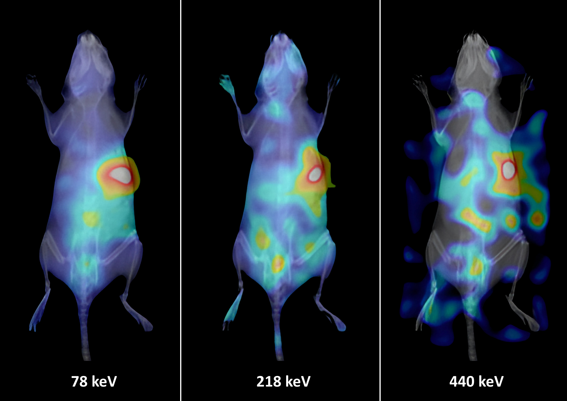
Figure 2: Planar imaging of a ²²⁵Ac-labelled FAPI-targeting agent in a HEK-FAP mouse model. Image info: 24 h post injection – 20 min scan duration – 0.19 MBq injected activity
²⁰³Pb and ²¹²Pb theranostic pair
The system was used to image the diagnostic and therapeutic members of a lead-based theranostic pair in a tumor-bearing mouse model, supporting comparative assessment of paired radiopharmaceutical behavior.
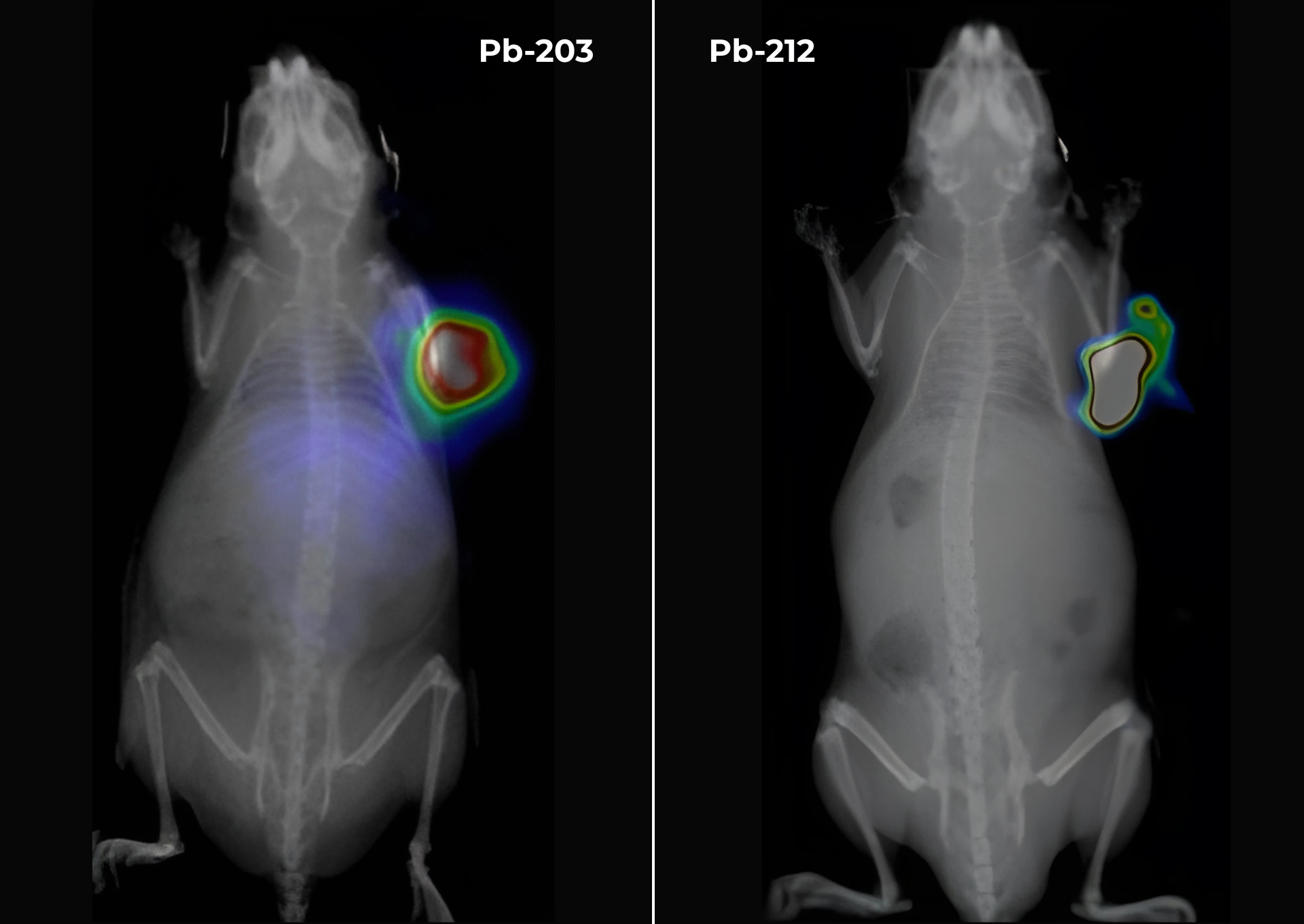
Figure 3: Planar imaging of a ²⁰³Pb and ²¹²Pb PSC-PEG-T theranostic pair. Image info: 24 h post injection – 10 min scan duration – injected activities: 0.89 MBq for ²⁰³Pb and 0.02 MBq for ²¹²Pb
Free ¹⁷⁷Lu distribution
Whole-body planar imaging enabled visualization of free ¹⁷⁷Lu distribution in a healthy mouse, including accumulation in the skeletal system, highlighting the value of planar imaging for straightforward whole-body distribution assessment.
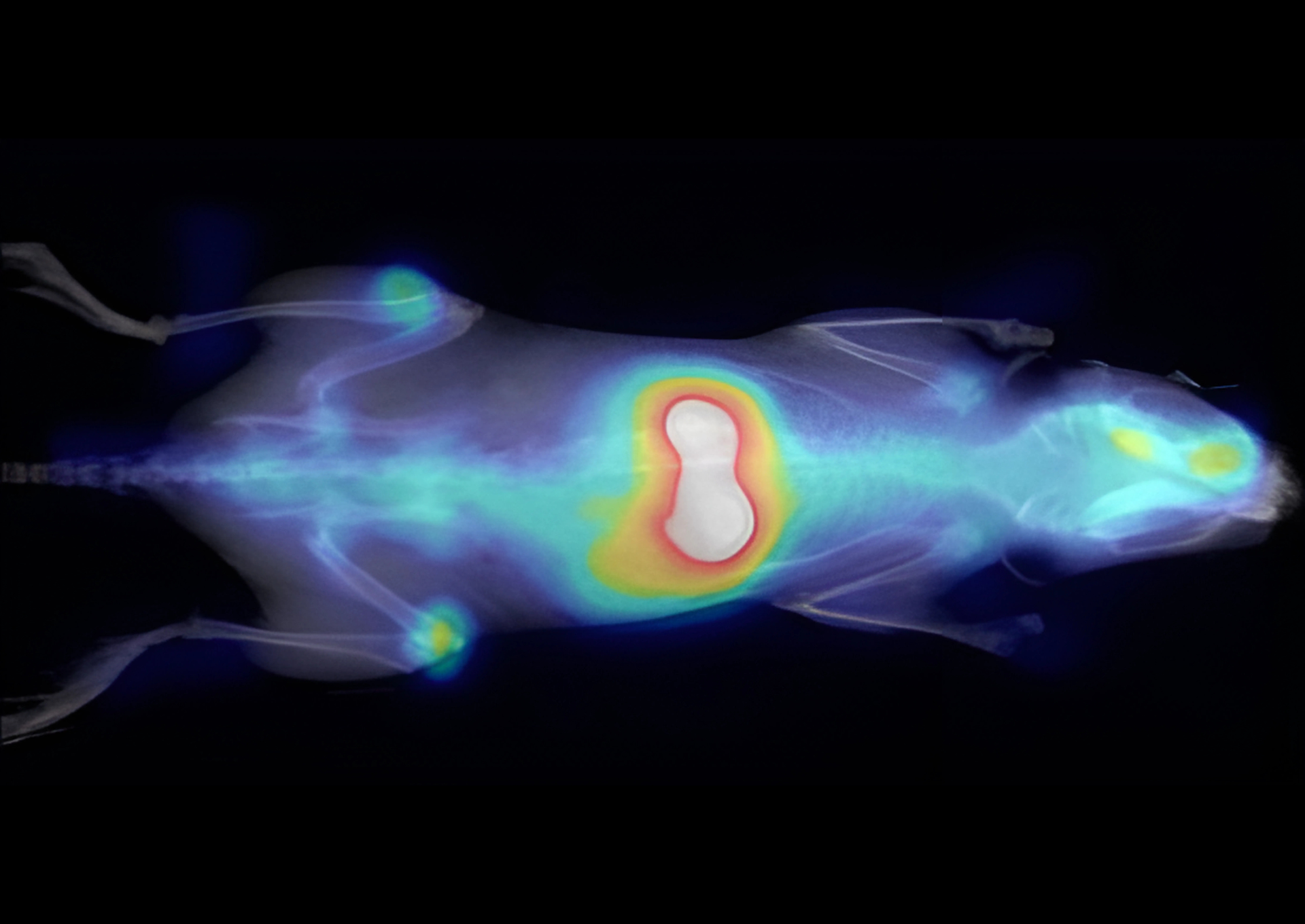
Figure 4: Planar imaging of free ¹⁷⁷Lu in a healthy mouse. Image info: 24 h post injection – 20 min scan duration – 20 MBq injected activity
¹¹¹In-DOTATATE tumor targeting
Planar imaging was used to assess the targeting properties of ¹¹¹In-DOTATATE under different experimental conditions, supporting rapid comparison of tracer uptake and distribution.
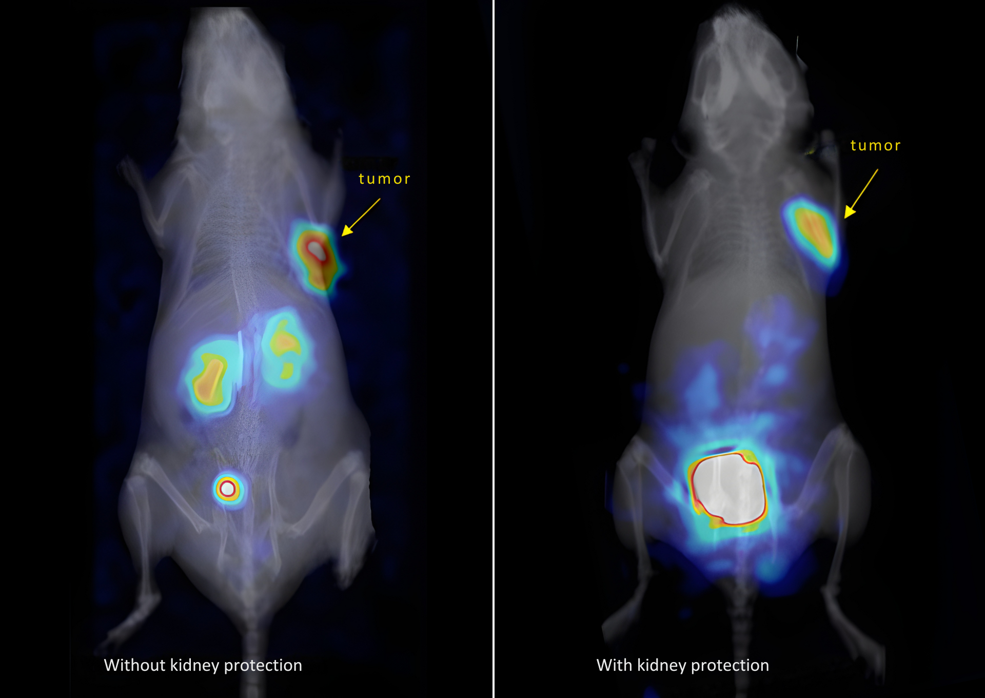
Figure 5: Planar imaging of ¹¹¹In-DOTATATE for assessment of tumor targeting, with and without tumor protection. Image info: 3 h post injection – 6 min scan duration – 4 MBq injected activity
Conclusion
These representative studies demonstrate the flexibility of γ-eye™ planar imaging across a broad range of radionuclides and radiopharmaceutical applications.
By enabling rapid whole-body visualization of tracer distribution, tumor targeting and early kinetic behavior, planar imaging extends the γ-eye™ workflow beyond tomographic SPECT and supports efficient in vivo screening, study optimization and selection of relevant follow-up imaging time points.