
Background
Actinium-225-labeled antibodies combine molecular targeting with high-linear-energy-transfer (high-LET) α emissions. Preclinical imaging remains difficult because administered activities are low and detectable photons arise mainly from daughter radionuclides in the ²²⁵Ac decay chain. Longitudinal multi-window SPECT/CT may provide serial whole-body distribution data within the same animal.
Objective
To evaluate longitudinal multi-window 3D SPECT/CT of [²²⁵Ac]Ac-DOTAGA-trastuzumab through 96 h under different count conditions, including late imaging following 55 kBq administered activity.
Radiopharmaceutical and imaging strategy
| Radiopharmaceutical | [²²⁵Ac]Ac-DOTAGA-trastuzumab |
| Model | HER2-positive SKOV-3 xenografts |
| Administered activity | 55 or 172 kBq |
| SPECT/CT | 30-min whole-animal acquisitions through 96 h |
| Reconstructions | 78, 218 and 440 keV; combined all-peaks |
| Terminal analysis | Ex vivo biodistribution at 96 h |
Study design
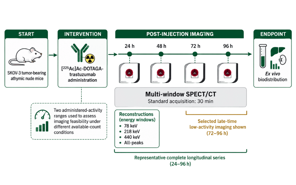
Radiochemistry and quality control
- Conjugation: DOTAGA (20 equiv.) + trastuzumab; overnight at 37 °C and 400 rpm, followed by 30-kDa Amicon purification and PBS washes.
- Radiolabeling: 0.5 M NH₄OAc (pH 5.9), target molar activity 28 kBq/nmol; 1 h at 37 °C and 400 rpm; reaction pH ~6.
- Formulation: 0.05% Tween-20 in 0.01 M PBS (pH 7.4).
- QC: iTLC-SG developed in 0.2 M citrate buffer (pH 4).
Labeled trastuzumab: 95.14% – Free ²²⁵Ac: 4.86%
Longitudinal imaging through 96 h
Representative complete longitudinal dataset after 172 kBq administered activity; 30 min per scan
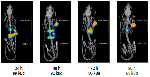
Figure 1: All-peaks SPECT/CT images from a complete longitudinal series. Values below the images show measured whole-body activity at acquisition.
Late-time whole-animal imaging following 55 kBq
Whole-animal coverage with 30-min acquisitions
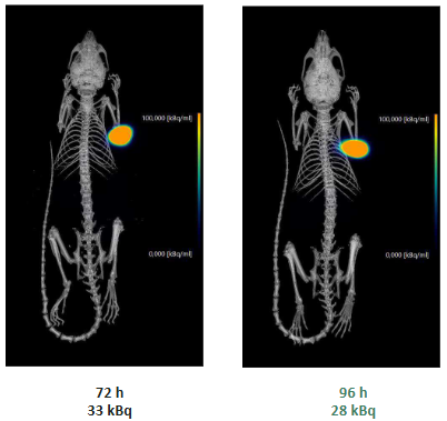
Figure 2: Representative late-time whole-body SPECT/CT after 55 kBq administered activity. Measured whole-body activity was 33 kBq at 72 h and 28 kBq at 96 h.
Decay-chain energy windows
Representative acquisition at 72 h after 172 kBq administered activity
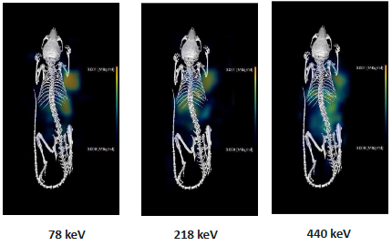
Figure 3: Window-specific SPECT/CT reconstructions.
Ex vivo biodistribution at 96 h
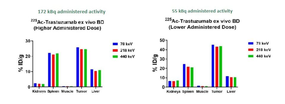
Figure 4: Representative ex vivo organ measurements at 96 h. Bars show estimates obtained independently from the 78, 218 and 440 keV measurements.
Key observations
- Persistent tumor-associated signal remained visible from 24 to 96 h in the complete longitudinal series.
- Whole-body acquisitions remained interpretable at 72 and 96 h when measured activity had decreased to 33 and 28 kBq, respectively.
- Window-specific reconstructions showed distinct spatial patterns while sampling emissions from the same decay chain.
- Terminal ex vivo organ measurements provided biological context for the SPECT findings.
Radiopharmacologic interpretation
Late-time count availability reflects the activity remaining in vivo after physical decay and biological clearance.
Whole-body SPECT/CT remained interpretable at 28 kBq measured activity at 96 h. Tumor-associated uptake remained prominent, with substantial spleen uptake. The plots describe each activity condition and do not constitute a formal comparison between administered activities.
Conclusions
- Longitudinal multi-window 3D SPECT/CT enabled non-invasive whole-body visualization of [²²⁵Ac]Ac-DOTAGA-trastuzumab through 96 h.
- Imaging remained interpretable at the final time point with 28 kBq measured whole-body activity following 55 kBq administered activity.
- Separate energy windows provided complementary information from the ²²⁵Ac decay chain.
- These findings support longitudinal preclinical radiopharmacology under reduced-count conditions.