Real time, small animal whole body imaging platform for fast screening of all PET radiopharmaceuticals
3D PET imaging
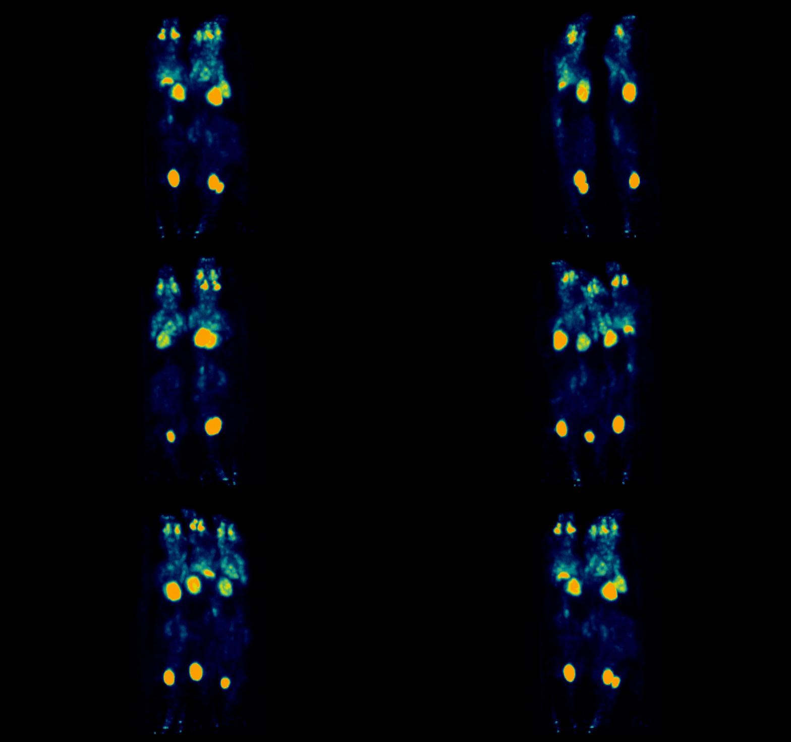
The β-eye™ system now features a 3D imaging option, expanding your capabilities at the press of a button.
- Simultaneous imaging of up to 3 mice, while maintaining effortless workflow and user-friendly operation
- Accelerated research throughput, with post-processing-ready data after just 6min of scanning & 1.5min of reconstruction
- Designed to deliver full-capability PET imaging in the smallest footprint on the market
- Upgrade your study portfolio with more applications like neuroimaging, orthotopic oncology and more
- Achieve enhanced quantification allowing comparisons across time points, subjects, and experimental conditions
- Supports dosimetry calculations in translational radionuclide therapy studies
- Distinguish and better understand complex structures
Features
- Small animal, PET isotope, 2D or 3D live imaging platform
- Real time, whole body imaging post injection
- Fast screening of radiolabeled compounds
- Suitable for organ ex vivo imaging
- Compatible with 3rd party anesthesia and vital signs monitoring systems

Definition of a user friendly platform
- Straightforward workflow with easy imaging protocol set-up
- Easy to operate by non highly-experienced professionals
- Footprint allowing for easy system installation and transfer
- Ideal for imaging inside a clean room, or next to a cyclotron
- Visual | eyes™ software hub for post processing of imaging data
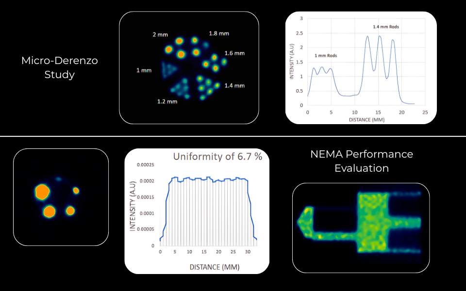
Performance
- State-of-the-art detector characteristics
- Spatial Resolution ~1mm
- Sensitivity of 2.9% @ Center of Field of View
- Contrast Recovery Coefficient (CRC) higher than 90% for up to 3mm rods
- Spill Over Ratio (SOR) of 11.7%
- Energy resolution of 12.4% @ 511keV
Applications
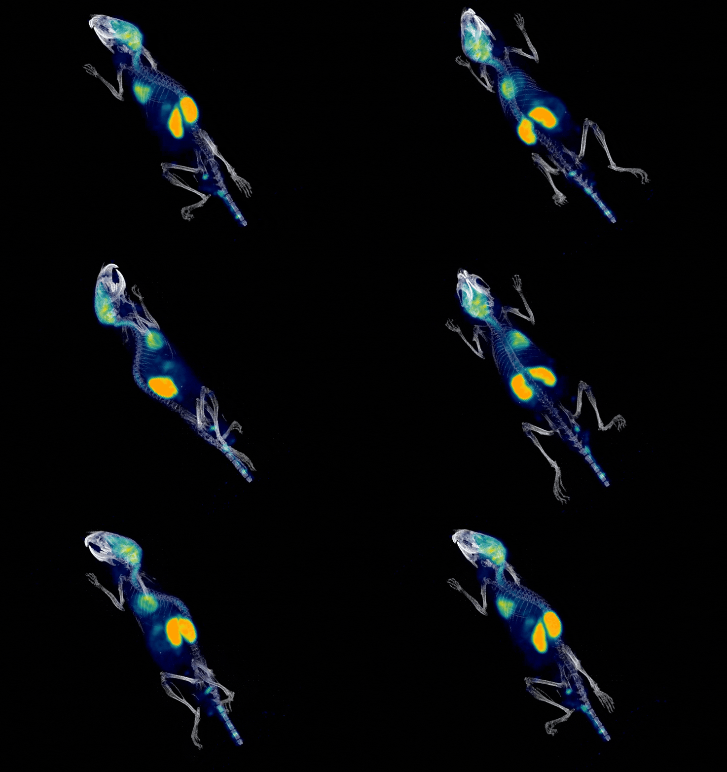
Brain imaging
Amyloid PET tracer kinetics
A preclinical PET application example supporting amyloid-tracer workflows
Image info: 2 min p.i. - 10 min scan duration - injected activity 2 MBq
Applications
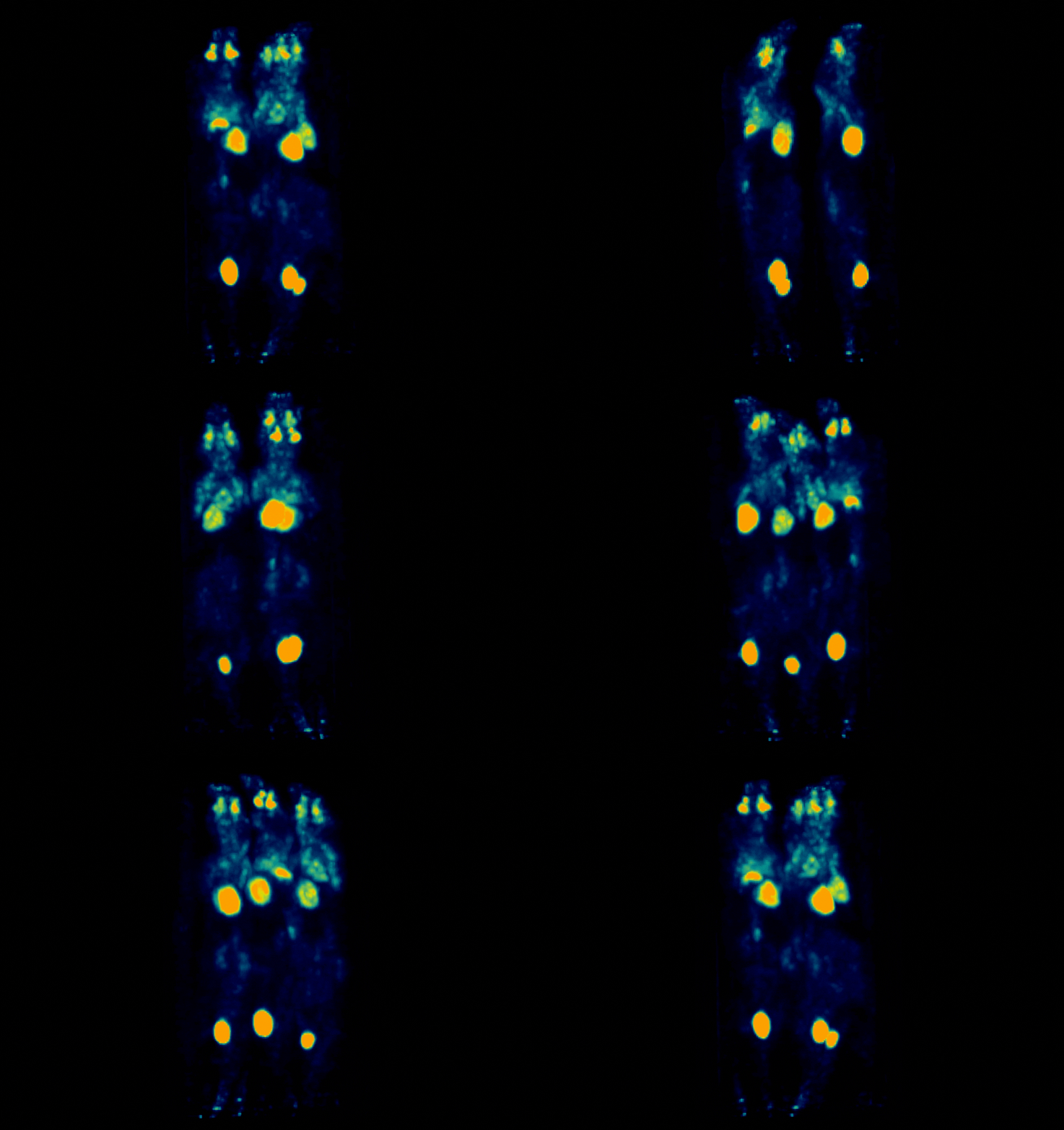
High-throughput PET imaging
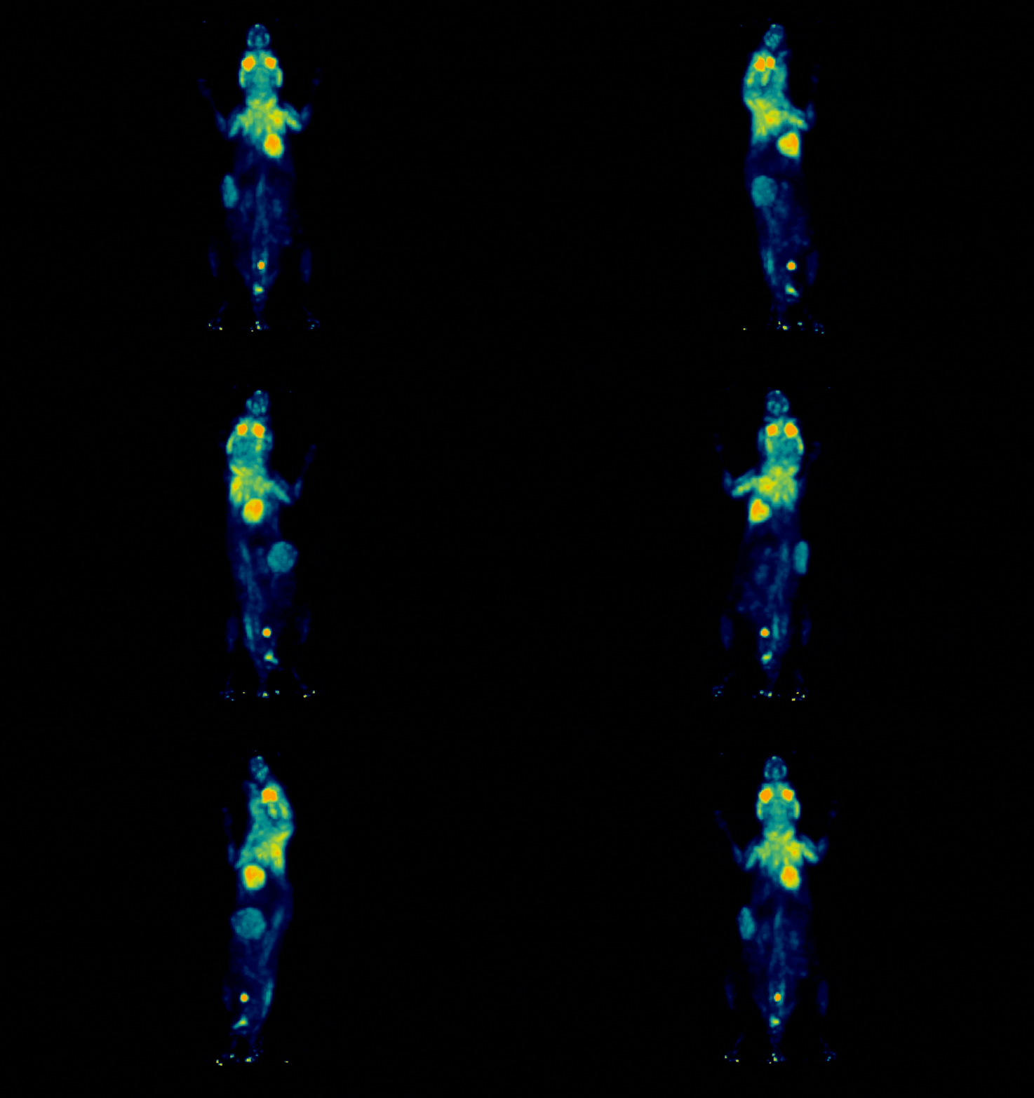
Simultaneous PET imaging of three mice
Image info: 70 min p.i. - 12 min scan duration - injected activity 2.34 MBq of ¹⁸F-FDG per mouse
Applications
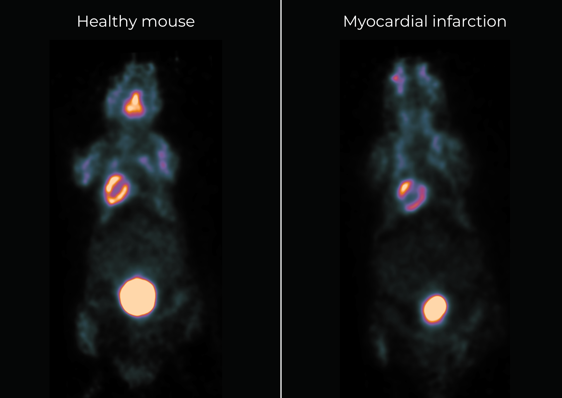
Cardiac PET
A preclinical platform to assess myocardial infarction therapies through non-invasive PET imaging
Cardiac PET imaging using ¹⁸F-FDG, in a mouse model of myocardial infarction
Image info: 80min post injection - 20min scan - total injected dose 6MBqApplications
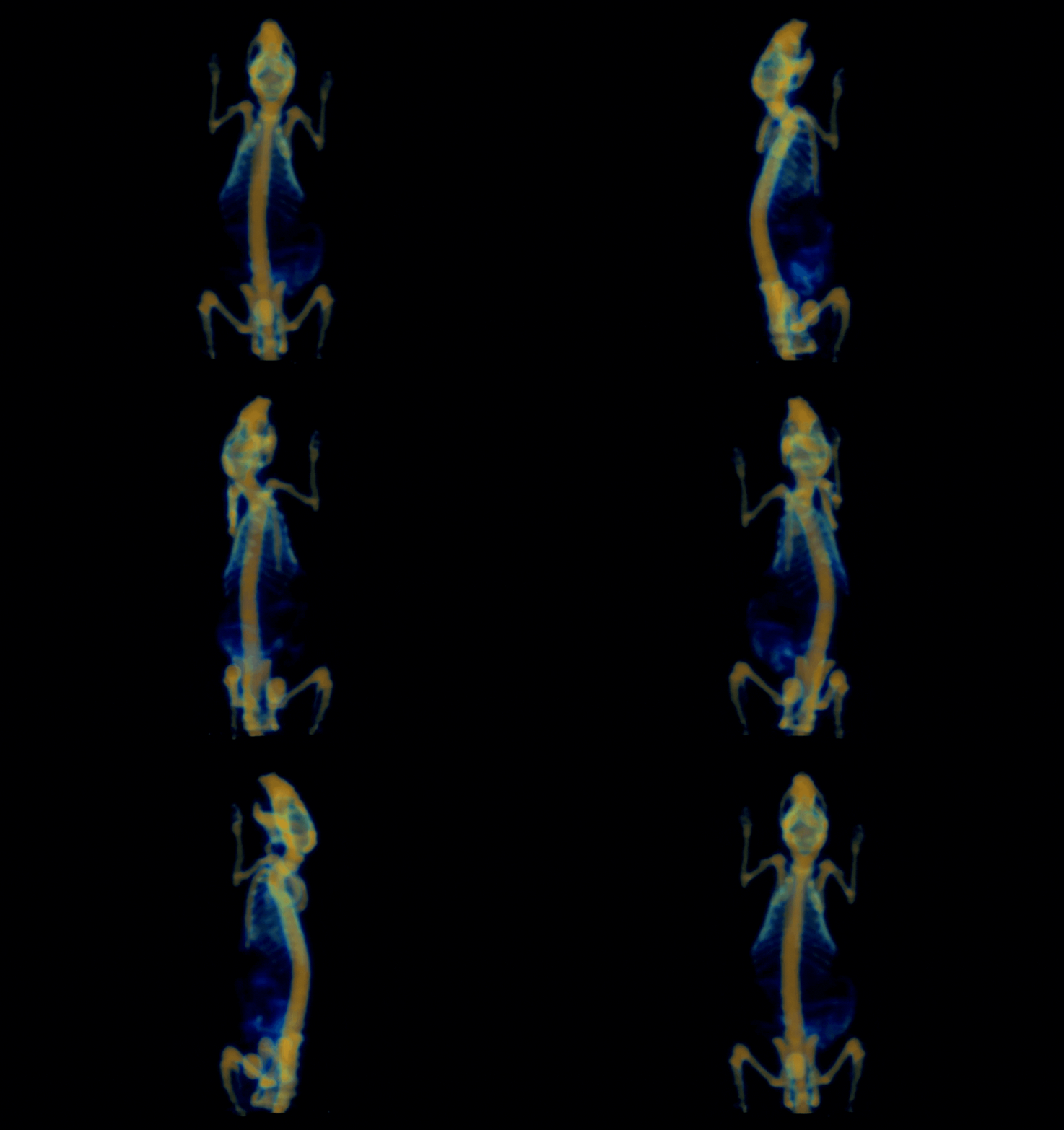
Bone imaging
Molecular tomography of skeletal structures
Distribution of free ¹⁸F in bone tissue
Image info: 1h post injection - injected activity 4MBqApplications
Oncology
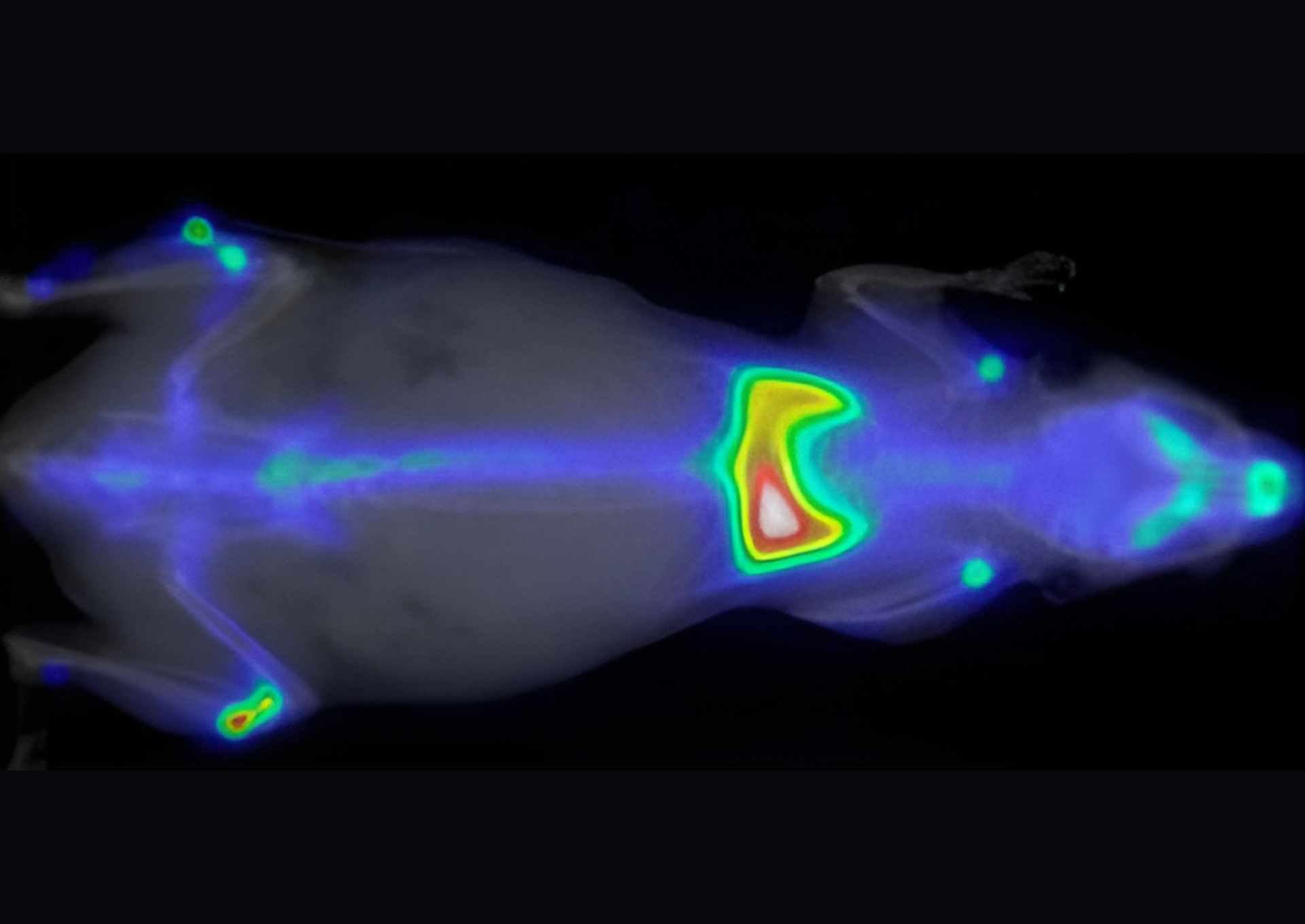
Metabolic phenotyping across tumor models using preclinical PET
Tumor model imaging using ¹⁸F-FDG
Image info: 1h post injection – 20 min scan - total injected activity 3MBqApplications
Planar imaging
Assessing pulmonary biology
Contamination model with ⁸⁹Zr to mimic real-world radiological exposure
Image info: 24h post administration - 5min scan - injected activity 3.7MBqApplications
Planar imaging
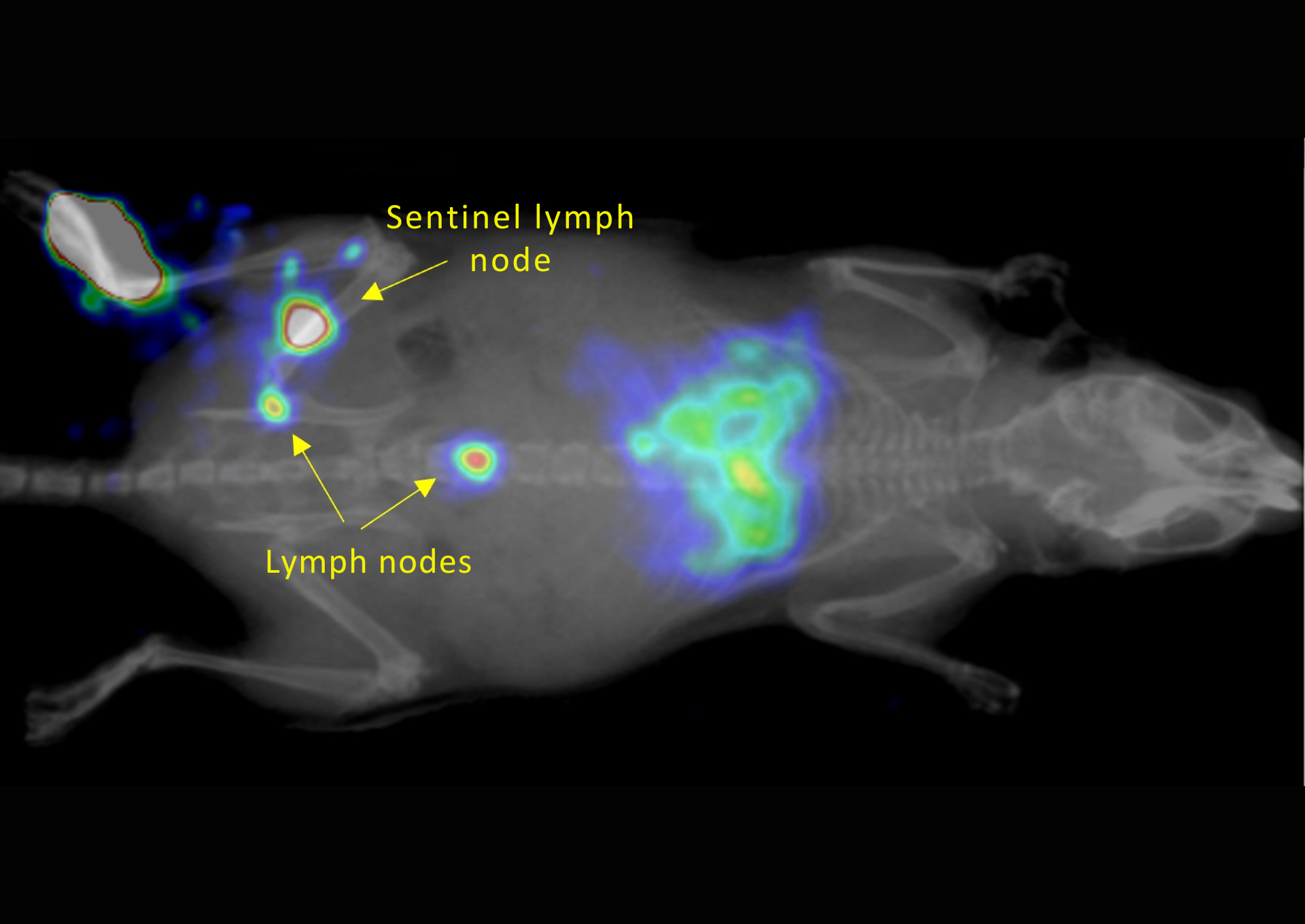
Preclinical imaging of lymphatic pathways
Sentinel lymph node imaging using ⁶⁸Ga-FAPI radiotracer
Image info: 1h post injection - 10min acquisition time - injected dose 3MBqApplications
Planar imaging
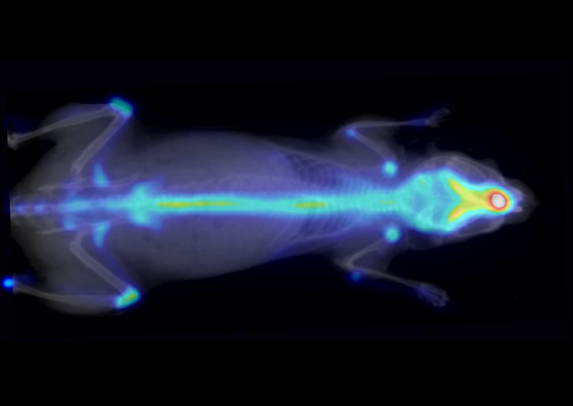
Assessing bone physiology
Free ¹⁸F accumulating in bones
Image info: 1h post injection acquisition - 10 min scan - injected dose 2.59 MBqApplications
Special applications
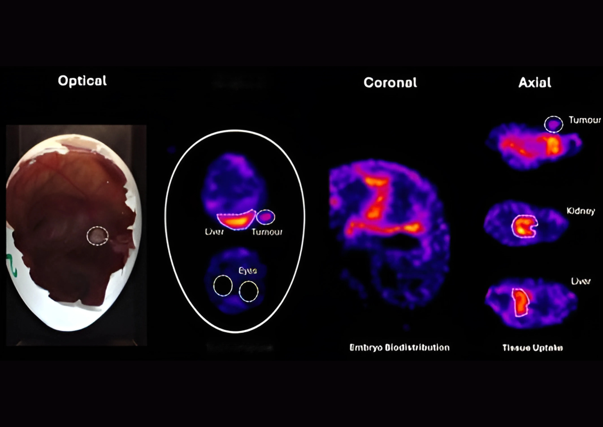
Imaging of embryonic models
In ovo imaging using ¹⁸F-based tracer
Images info: 2h post injection - 10 min scanApplications
Special applications
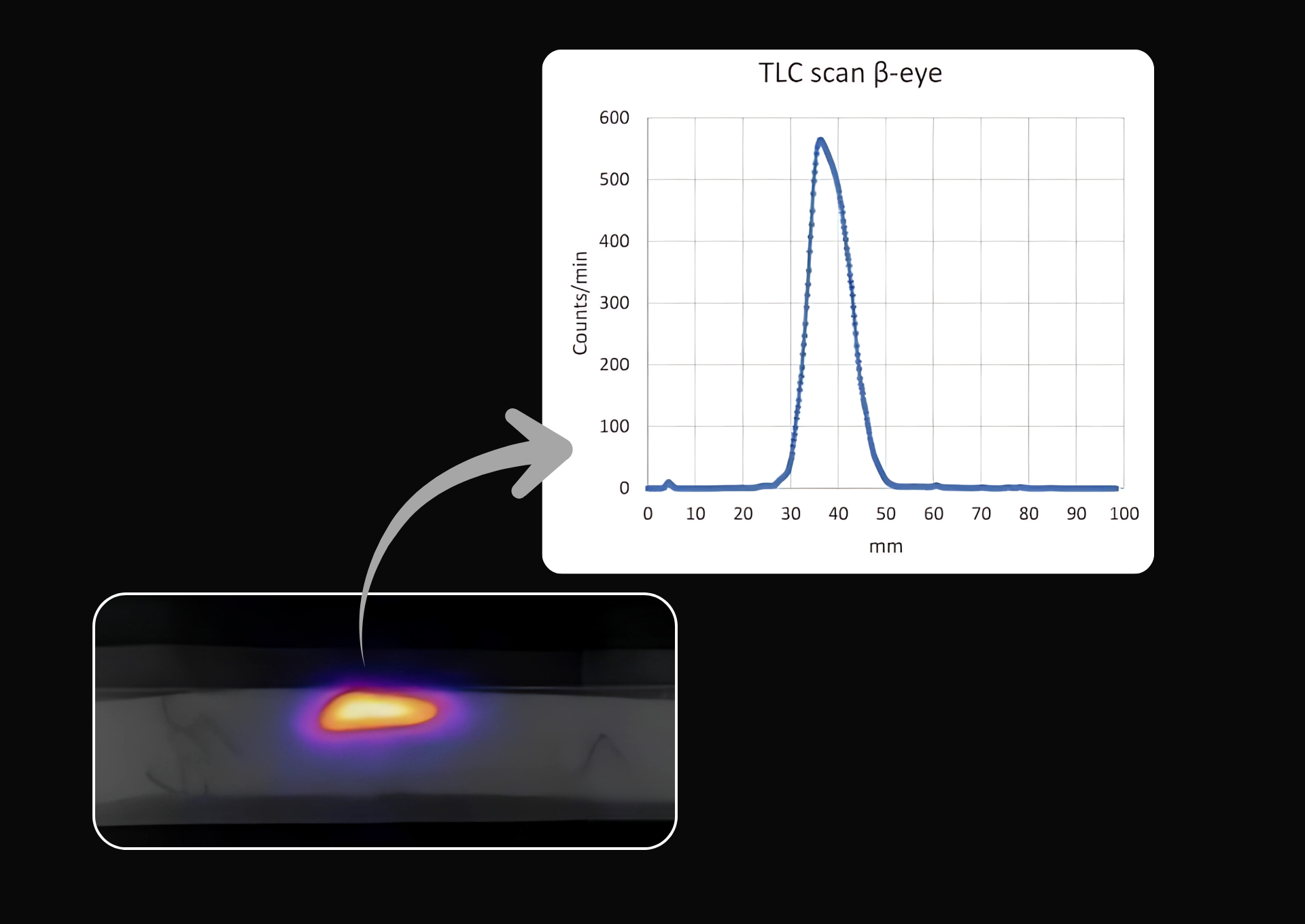
TLC scan
Thin-layer chromatography of ⁶⁴Cu-labeled compound
Image info: 3 min scan - injected activity 0.26 MBq
Publications
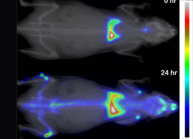 A Murine Model of Radionuclide Lung Contamination for the Evaluation of Americium Decorporation TreatmentsRead More
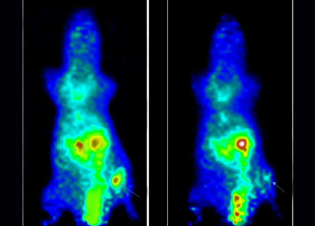 Preparation and preclinical evaluation of 18F-labeled folate-RGD peptide conjugate for PET imaging of triple-negative breast carcinomaRead More
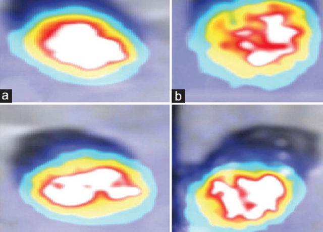 Cardioprotective effect of S-adenosyl L-methionine due to antioxidant and anti-inflammatory properties on isoproterenol-induced chronic heart failure in Wistar ratsRead More
Expert insights on preclinical PET imaging
A recent webinar by a member of the BIOEMTECH team explores key principles and real-world examples of preclinical metabolic PET imaging using the β-eye™ system.

