Highly sensitive, desktop, in vivo optical imaging system for Fluorescence and Bioluminescence preclinical studies

Features
- Small animal, whole body, live optical-imaging platform
- Fluorescence and Bioluminescence – Suitable for Cherenkov imaging (CLI) studies
- Extension to SWIR imaging
- Up to 5 mice simultaneous imaging
- Compatible with 3rd party anesthesia and vital signs monitoring systems

Performance
- High Quantum Efficiency of 85% at 460nm / 70% at 600 nm / 30% at 900 nm
- Dark current down to 0,016 e/pixels/sec
- Ultimate performance for low-signal applications
- Resolution of 4,6 μpixel – 4096 x 2304
- Readout noise as low as 0,3 electrons (rms)
- Photon number resolving imaging / Quantitative CMOS
- Weight less than 70kg
- Dimensions: 60cm (L) x 53cm (W) x 60cm (H)

Definition of a user friendly platform
- Straightforward workflow with easy imaging protocol set-up
- Plain to operate by non highly-experienced professionals
- User exchangeable optical filters thanks to BIOEMTECH’s unique filter cassettes concept
- Footprint allowing for simple system installation and transfer
- Ideal for imaging inside a clean room
- Visual | eyes™ software hub for post processing of imaging data
Applications
Oncology, BLI
Monitoring tumor growth with Bioluminescence imaging
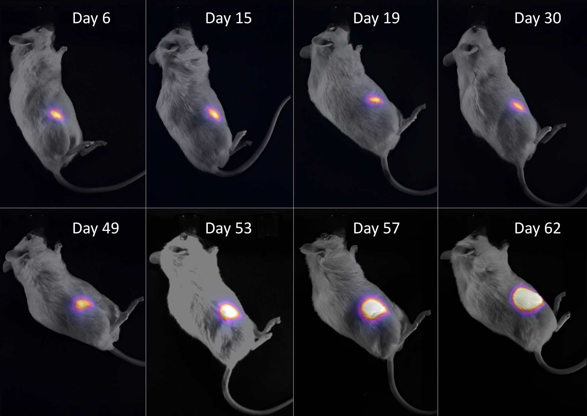
Α subcutaneous U87MG_Luc2 tumor was monitored over 62 days. Bioluminescence imaging (BLI) with D-luciferin revealed stable signals during the early phase, followed by a sharp increase after day 49, reflecting accelerated tumor growth.
Applications
Oncology, FLI
Evaluation and staging of Epidermoid Carcinoma
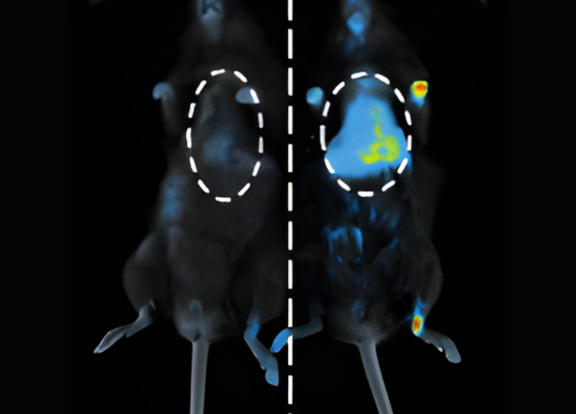
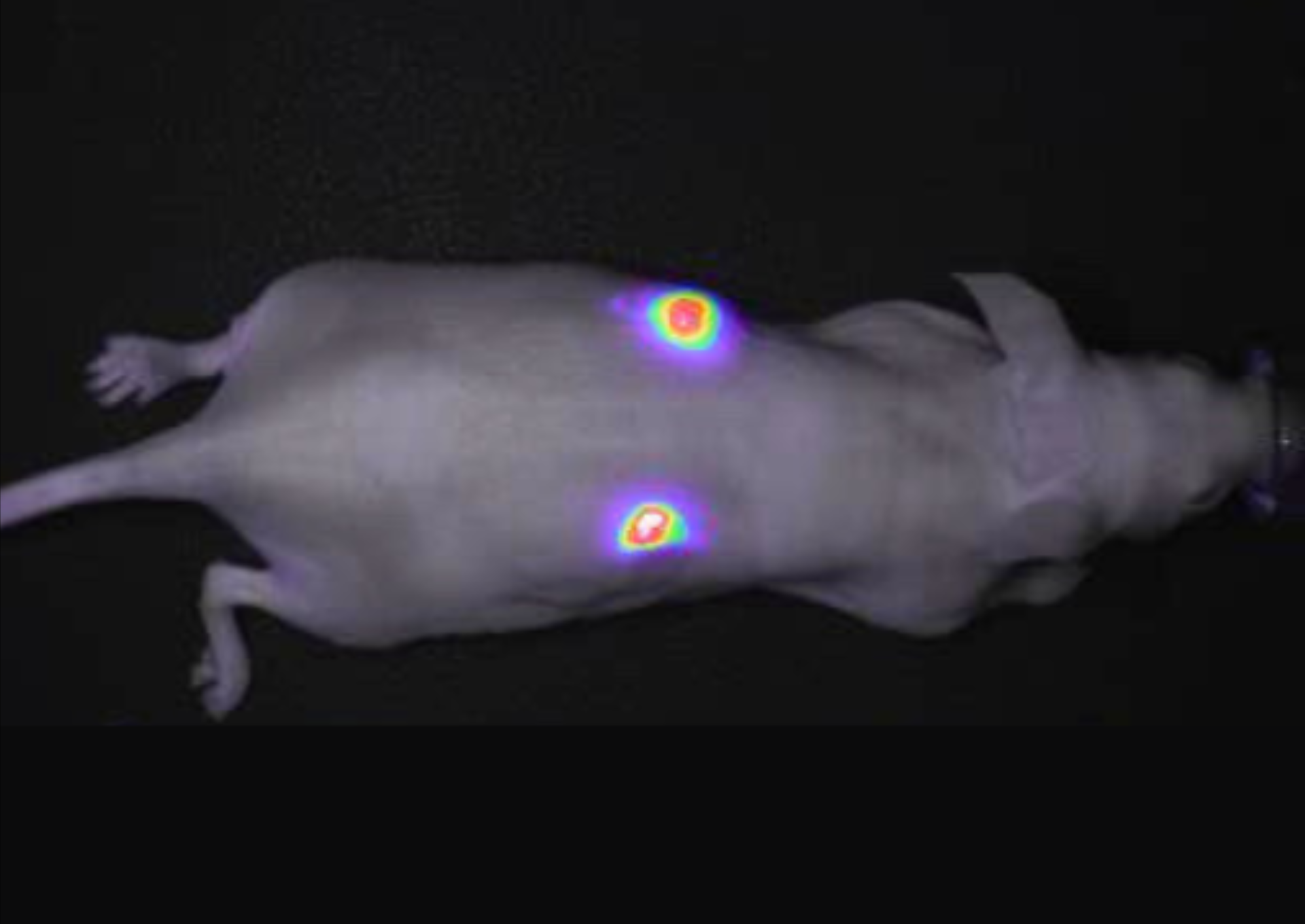
This case study highlights the use of fluorescence optical imaging for in vivo therapy evaluation and staging of Epidermoid Carcinoma (Squamous Cell Carcinoma) in preclinical models. RFP dyes were utilized to enable targeted imaging and enhance visualization of tumor dynamics.
Applications
Oncology, FLI
Tumor imaging using the φ-eye™
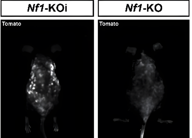
tdTomato, a constitutively fluorescent orange protein (excitation ~554 nm, emission ~581 nm), is utilized for tumor imaging applications. This example demonstrates its use in a neurofibroma model, with the dye administered intravenously to enable fluorescence-guided imaging.
Applications
SWIR, FLI
Imaging healthy mice in the SWIR window
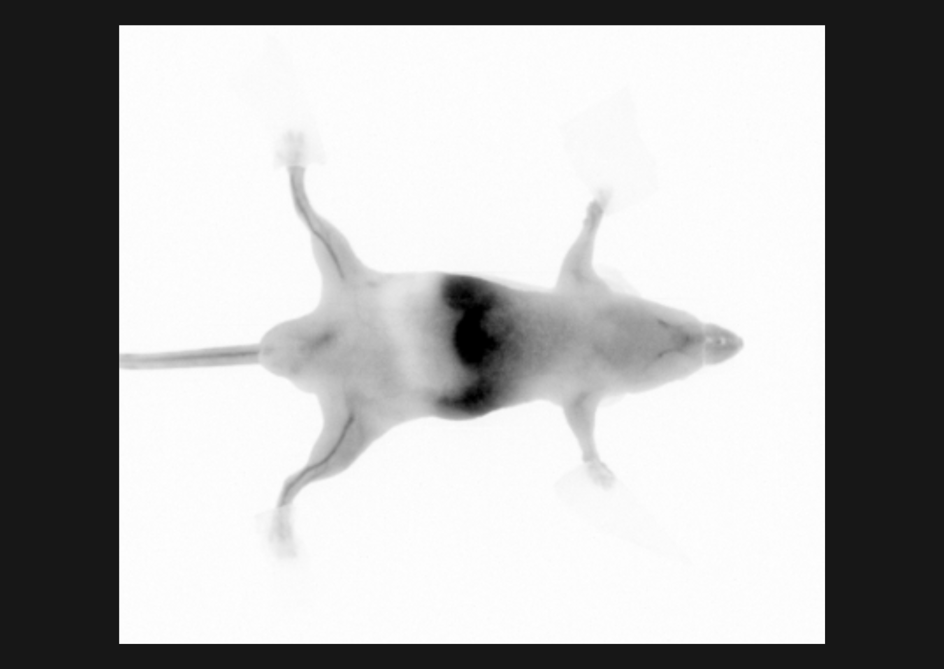
BIOEMTECH's technology enables imaging in the Short Wavelength InfraRed (SWIR) window. This example demonstrates clear visualization of the saphenous vein in a healthy mouse. Imaging parameters: Indocyanine Green (ICG) dye, injected volume 100 μL, concentration 500 μM, 1 min post-injection (p.i.) and 5 ms exposure time.
Applications
Inflammation, FLI
Bone imaging using the φ-eye™
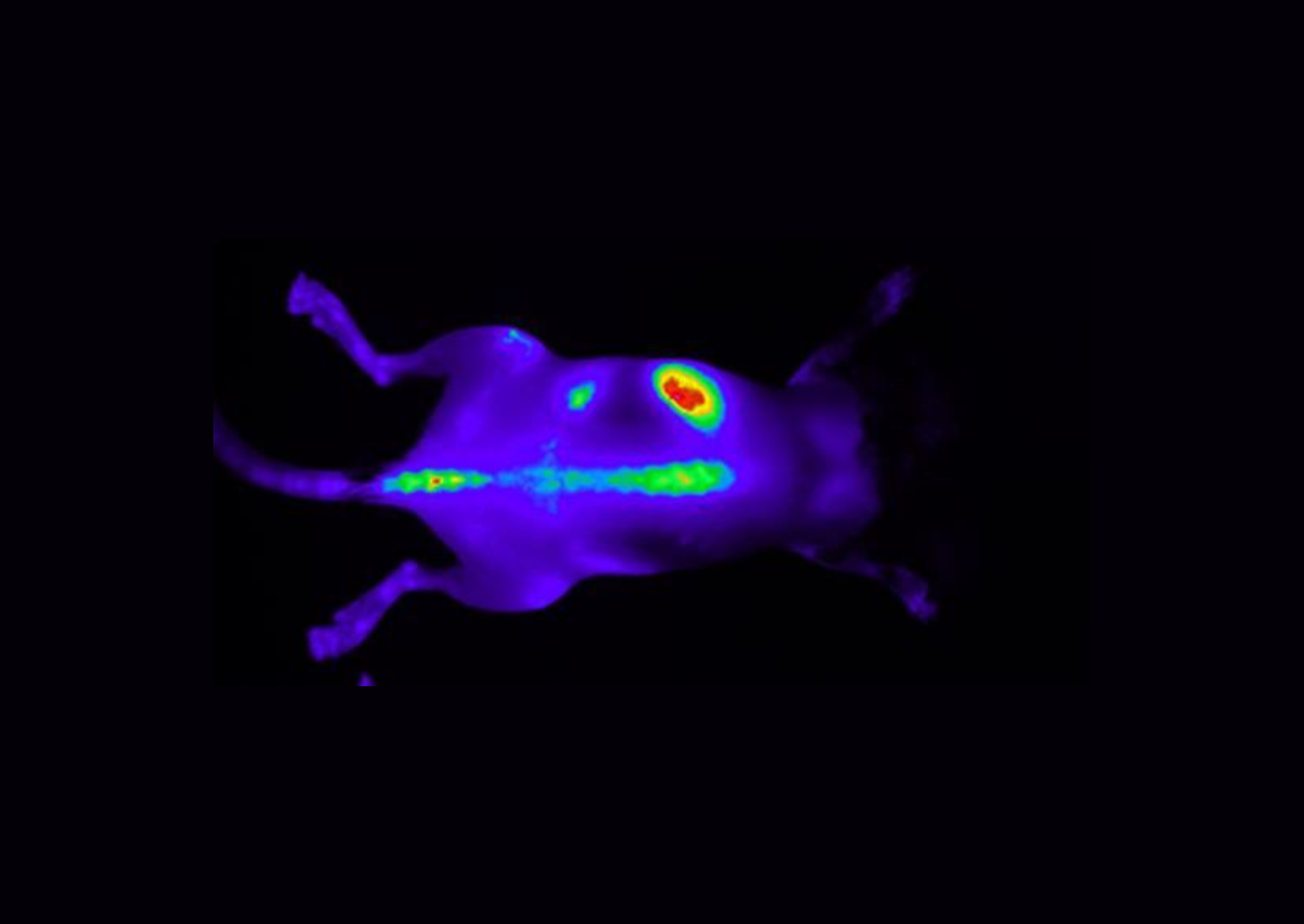
In vivo fluorescence imaging of bone was performed using a probe with an emission wavelength of 680 nm in a mouse model of spondyloarthritis. This approach offers a powerful tool for monitoring disease progression and evaluating the efficacy of novel therapeutic interventions.
Applications
Oncology, BLI
Tumor imaging using a bioluminescent substrate
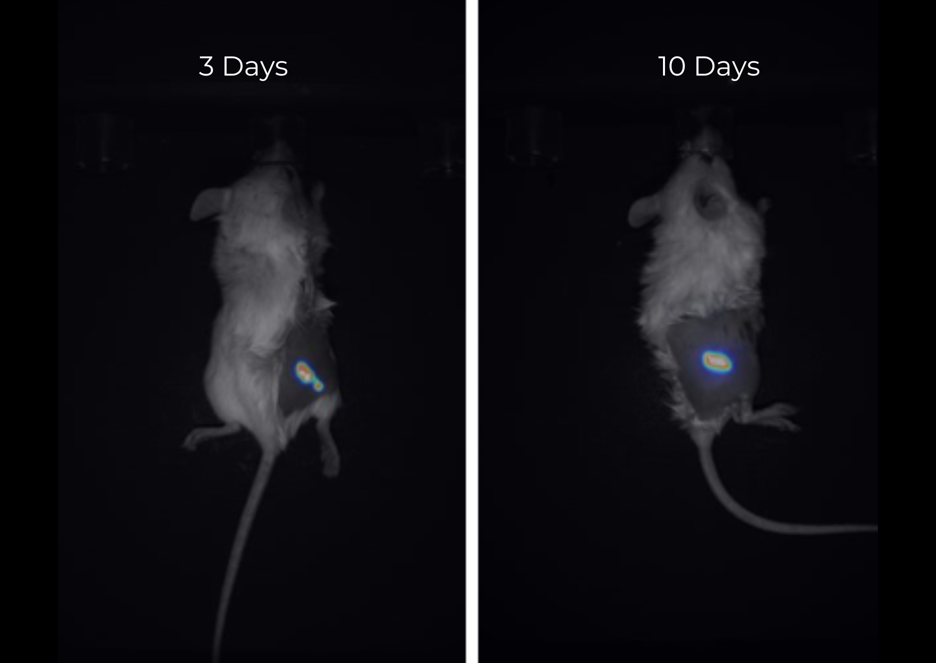
This study demonstrates the use of D-Luciferin as a substrate for bioluminescence imaging in a non-orthotopic glioblastoma tumor model. Imaging was performed at day 3 and 10 after the inoculation of 1 million U87MG-Luc2 cells. Key parameters: injected concentration of 3 mg, injection volume of 200 μL saline and exposure time of 5 minutes.