
Real-time, in vivo rodent imaging, allows researchers to observe and monitor several normal and abnormal biological processes. Data collected using the eyes™ series, provides crucial initial insights and generate quantitative results that, in combination with the Visual | eyes™ interface, compose an essential platform for every preclinical laboratory.
Oncology
Non-invasively assess your agent's targeting properties, real-time monitor drug delivery and track tumor progression.
Infection, Inflammation
Acquire detailed insights into disease mechanisms, enhance more accurate diagnostics while supporting the development or improvement of new or already existing therapeutics.
Protocol optimization
Test different animal preparation conditions, drug concentrations and administration routes to optimize your protocols.
Quick insights
Scan the same mouse at multiple time points to determine the optimal moments for 3D imaging. Conduct super-fast QC to exclude faulty injected subjects from further and more complex studies.
Applications with eyes™
Oncology, BLI
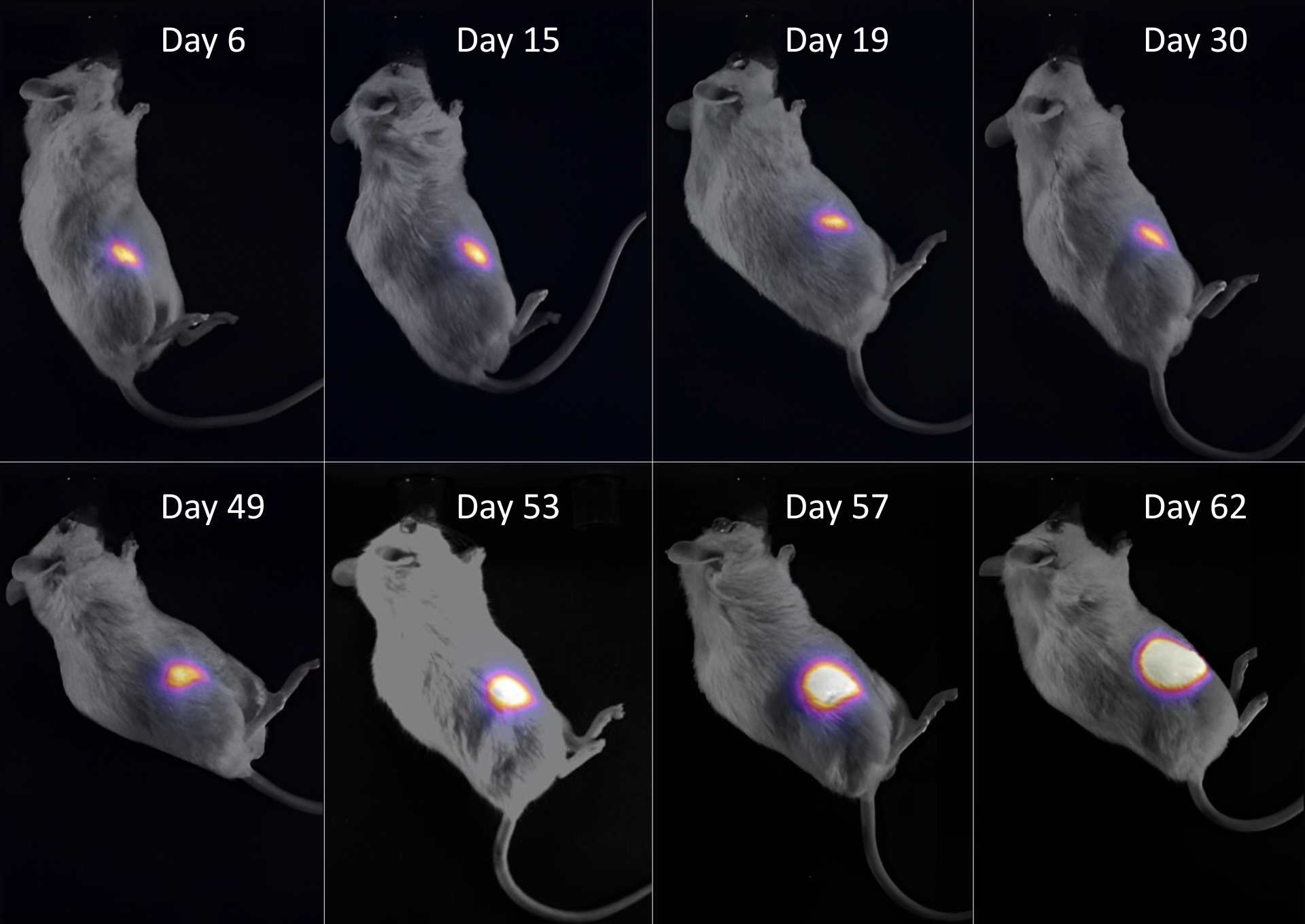
Bridging 𝑰𝒏 𝑽𝒊𝒕𝒓𝒐 and 𝑰𝒏 𝑽𝒊𝒗𝒐: Longitudinal Imaging of Glioblastoma Growth Across Experimental Scales
Α subcutaneous U87MG_Luc2 tumor was monitored over 62 days. Bioluminescence imaging (BLI) with D-luciferin revealed stable signals during the early phase, followed by a sharp increase after day 49, reflecting accelerated tumor growth.
Study performed by the LBMI group at FORTH-IESL and presented at EMIM 2026.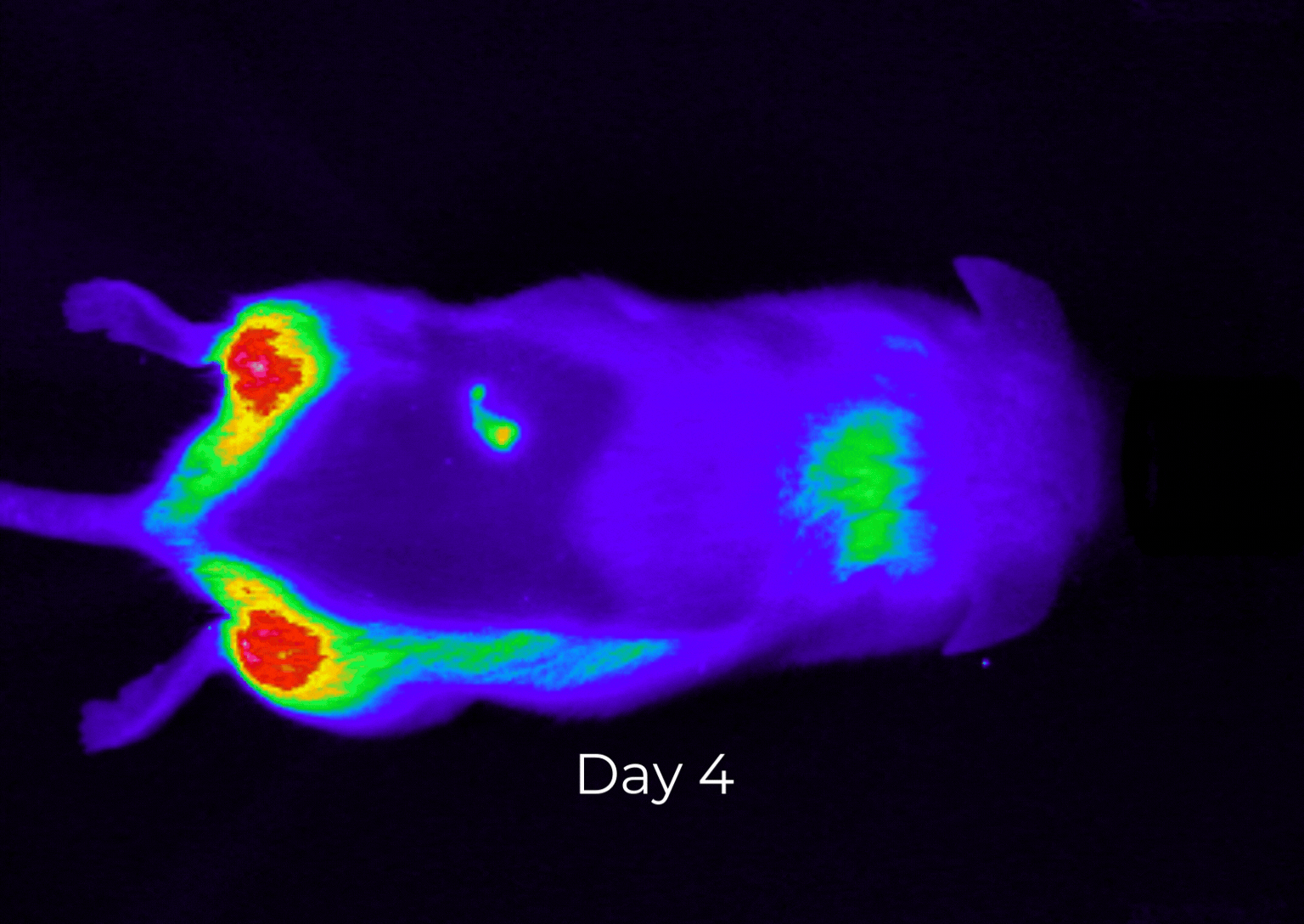
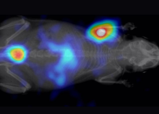
Oncology, FLI
Evaluation and staging of Epidermoid Carcinoma
This case study highlights the use of fluorescence optical imaging for in vivo therapy evaluation and staging of Epidermoid Carcinoma (Squamous Cell Carcinoma) in preclinical models. RFP dyes were utilized to enable targeted imaging and enhance visualization of tumor dynamics.
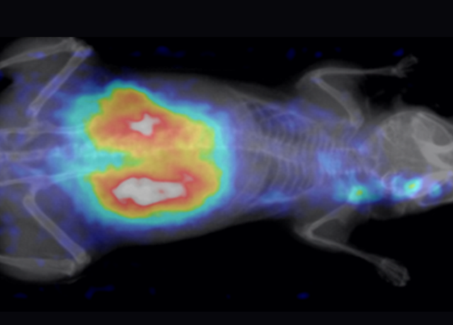
Oncology, FLI
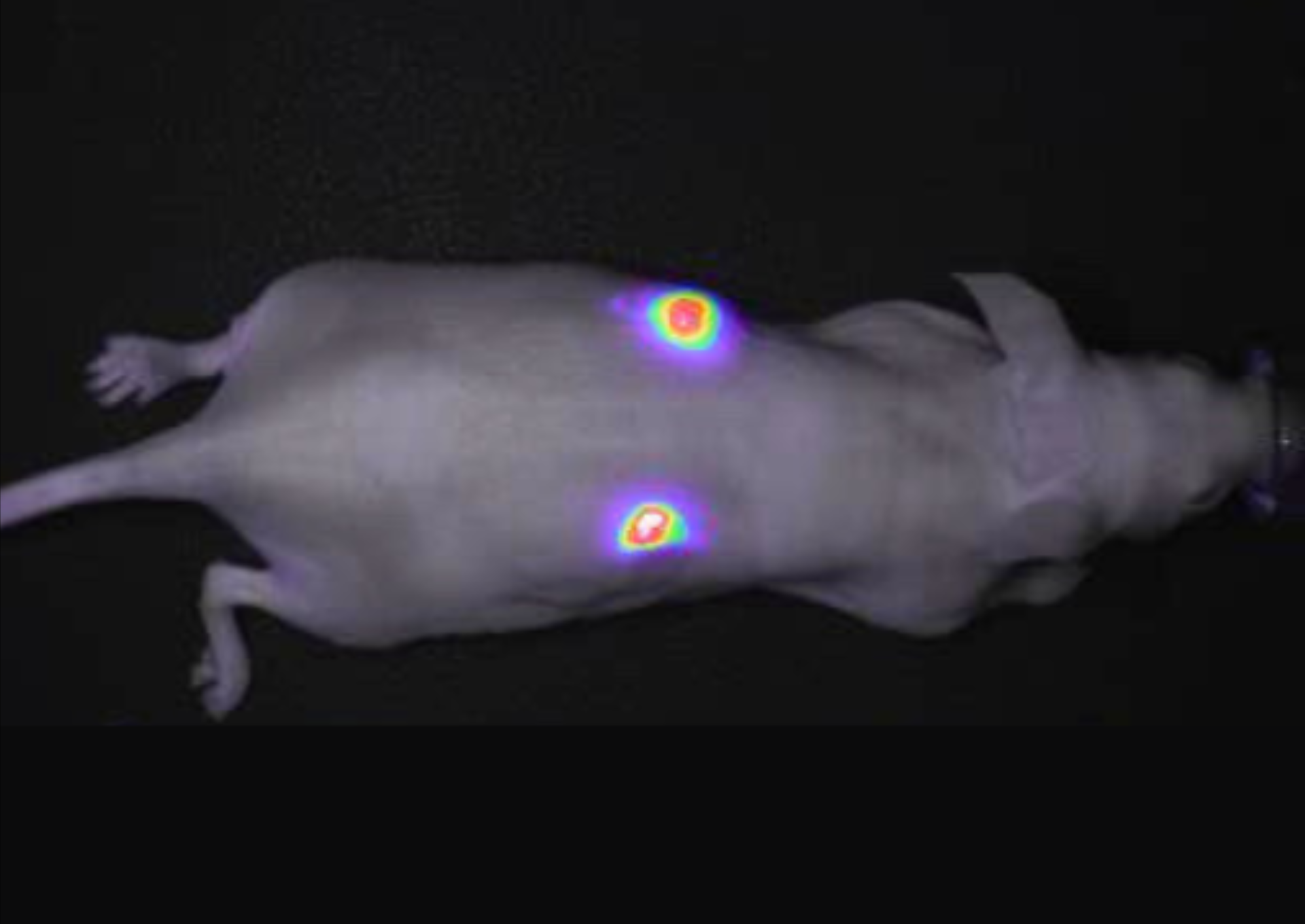
Tumor imaging using the φ-eye™
tdTomato, a constitutively fluorescent orange protein (excitation ~554 nm, emission ~581 nm), is utilized for tumor imaging applications. This example demonstrates its use in a neurofibroma model, with the dye administered intravenously to enable fluorescence-guided imaging.