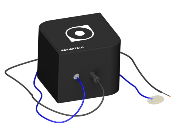
“Embracing scientists translate ideas into outcomes.”
…by providing the right tools for fast and effective research: meet the eyes™ series.
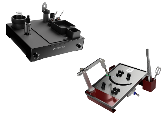
Products
Applications
Oncology, alpha emitters
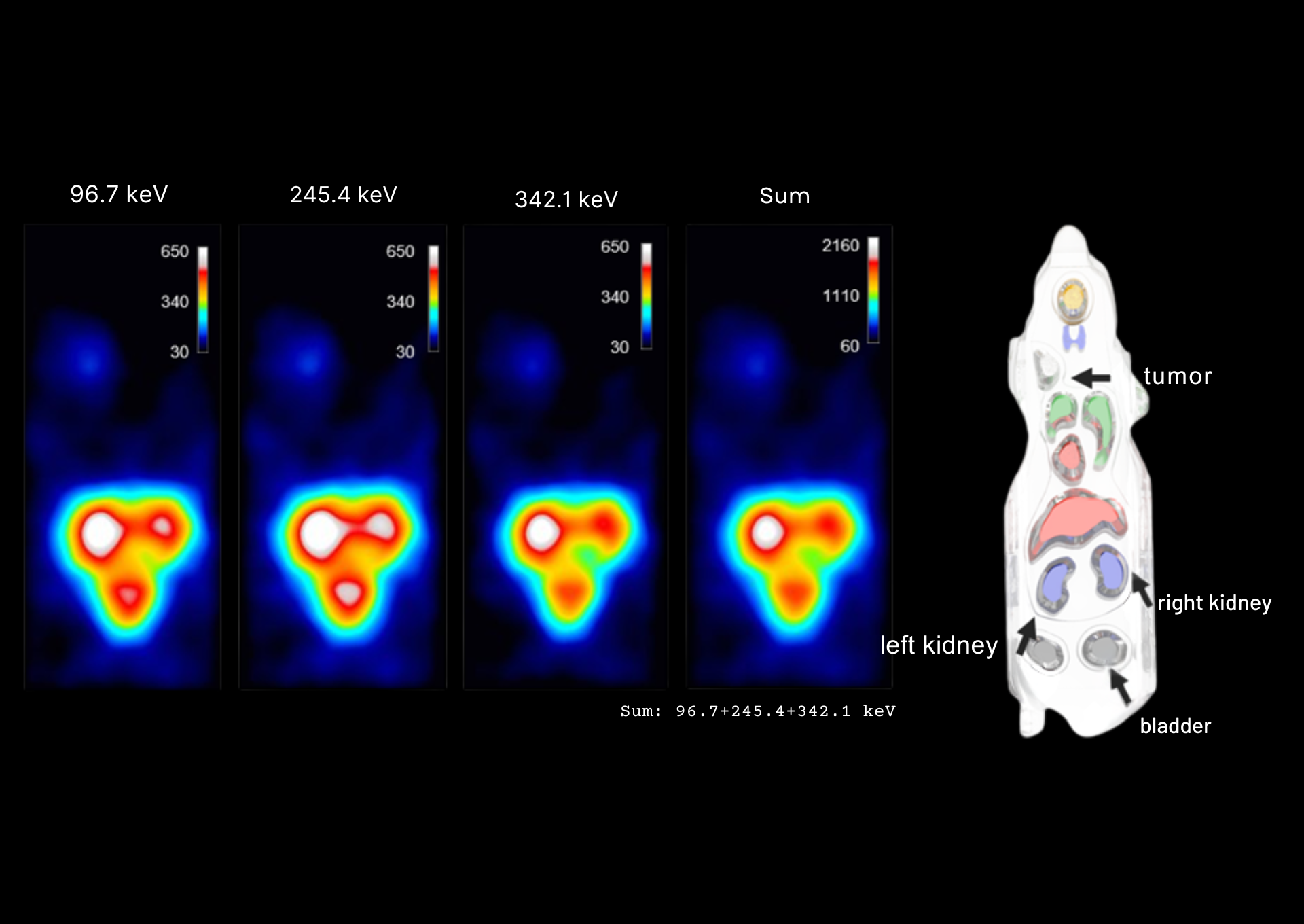
3D SPECT imaging across the ²²⁵Ac decay chain
Multi-window imaging of ²²⁵Ac-Trastuzumab in a SKOV3 tumor model
Image info: 48 h post injection - 30 min scan duration - 172kBq injected activity
Study performed in BIOEMTECH Laboratories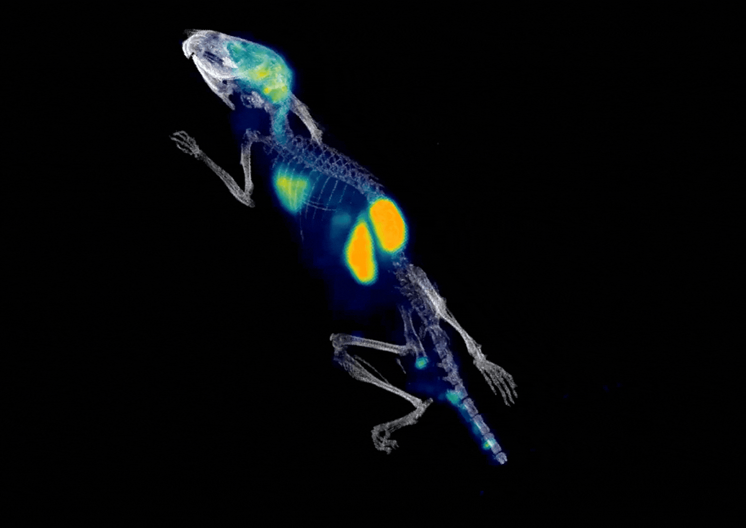
Applications
Brain imaging
Amyloid PET tracer kinetics
A preclinical PET application example supporting amyloid-tracer workflows
Image info: 2 min p.i. - 10 min scan duration - injected activity 2 MBq
Applications
High-throughput PET imaging
Simultaneous PET imaging of three mice
Image info: 70 min p.i. - 12 min scan duration - injected activity 2.34 MBq of ¹⁸F-FDG per mouse
Applications
Theranostics
Evaluation of preclinical quantitative imaging of ¹¹¹Ag
Optimization and validation in a mouse phantom
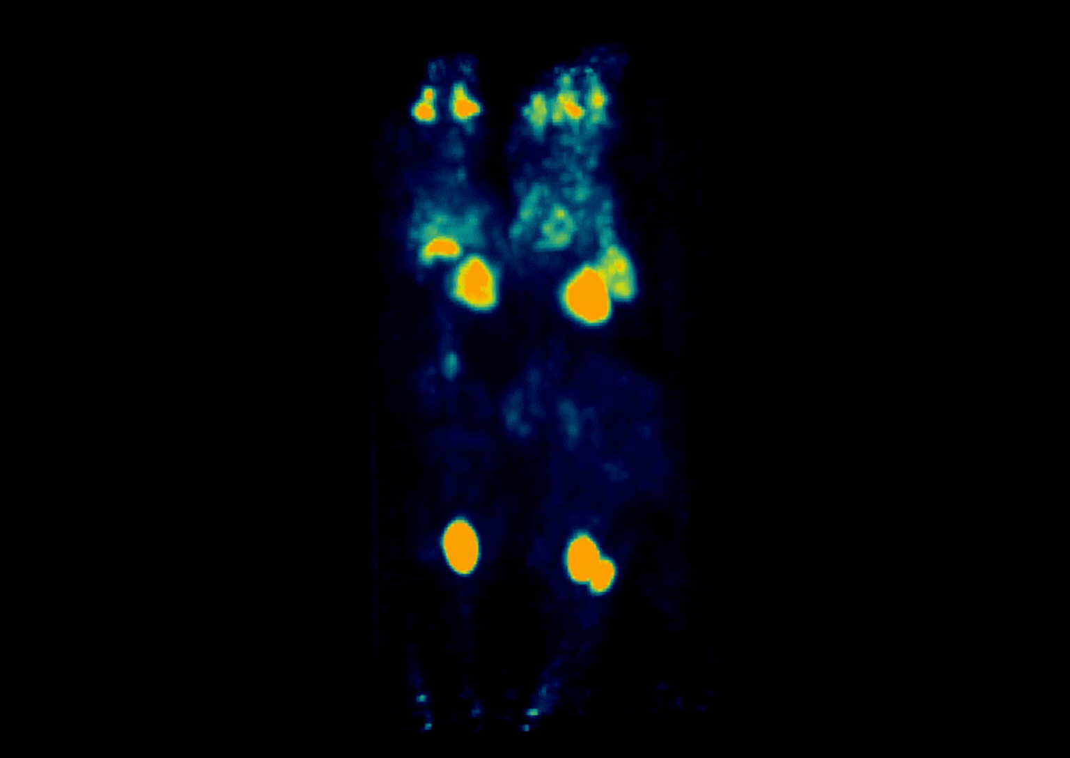
Applications
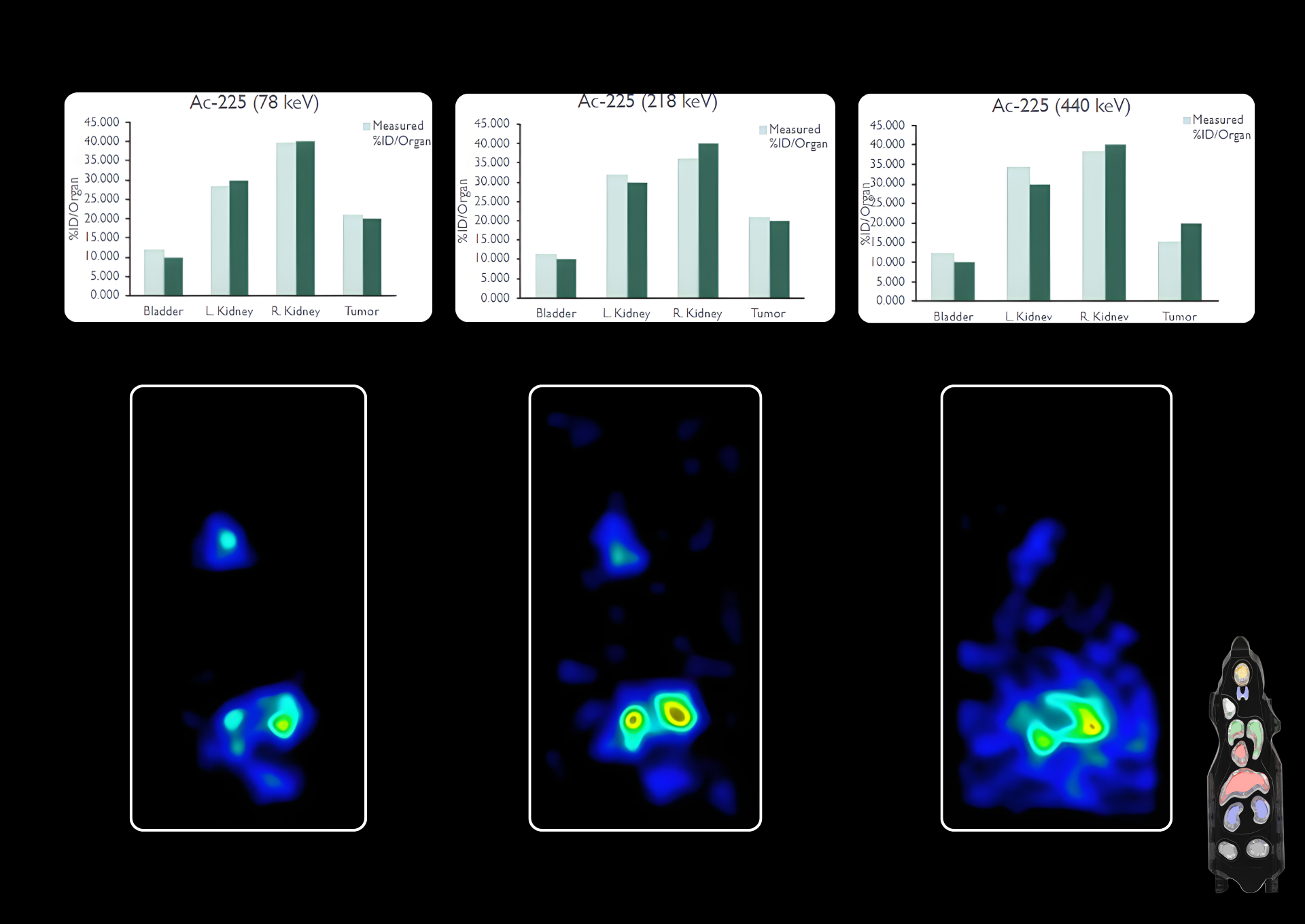
Alpha-emitters
Quantification phantom studies
Phantom imaging with ²²⁵Ac and quantification plots, acquired at 3 characteristic energy peaks of the isotope